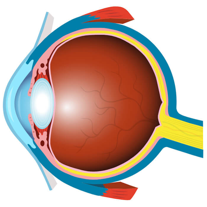
Anatomy of the eye
Click on the buttons to learn about the functionality of the optic nerve, pupil, conjunctiva and other parts of the eye.
Cornea
The clear, dome-shaped front part of the eye that helps your eye focus light, so you can see clearly.
Pupil
The opening in the centre of the iris that controls the amount of light passing through to the back of the eye.
Crystalline Lens
The transparent, elastic structure inside the eye that bends to focus light rays onto the retina.
Aqueous Humour
The clear, watery fluid in the front of the eyeball made up of water, sugars and various nutrients. It nourishes the cornea and the lens and gives the eye its shape.
Iris
The coloured part of the eye surrounding the pupil that controls the amount of light that enters into the eye.
Conjunctiva
A clear, thin transparent layer of tissue that covers part of the front surface of the eye and the inner surface of the eyelids.
Sclera
The dense protective tissue of the eyeball that forms the ‘white’ of your eye. Sclera forms over 80% of the surface area of the eyeball, from the cornea to the optic nerve.
Vitreous Humour
A clear gel-like substance that occupies the space between the crystalline lens and the retina and transmits initial light waves.
Optic Disc (Blind Spot)
A disc on the retina that is the point of entry of the optic nerve. It lacks visual receptors, so it’s also known as the blind spot.
Macula
The central part of the retina that allows us to see fine details.
Fovea
The central part of the macula, necessary for activities where visual detail is important, such as driving or reading.
Retina
The light-sensitive nerve layer that lines the inside of the back of the eye. It creates electrical signals that travel through the optic nerve to the brain.
Optic nerve
A bundle of nerve fibres that connect the retina with the brain and help us interpret what we see.